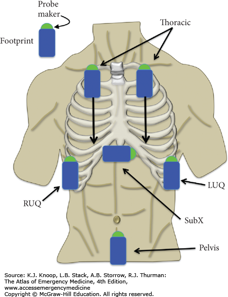
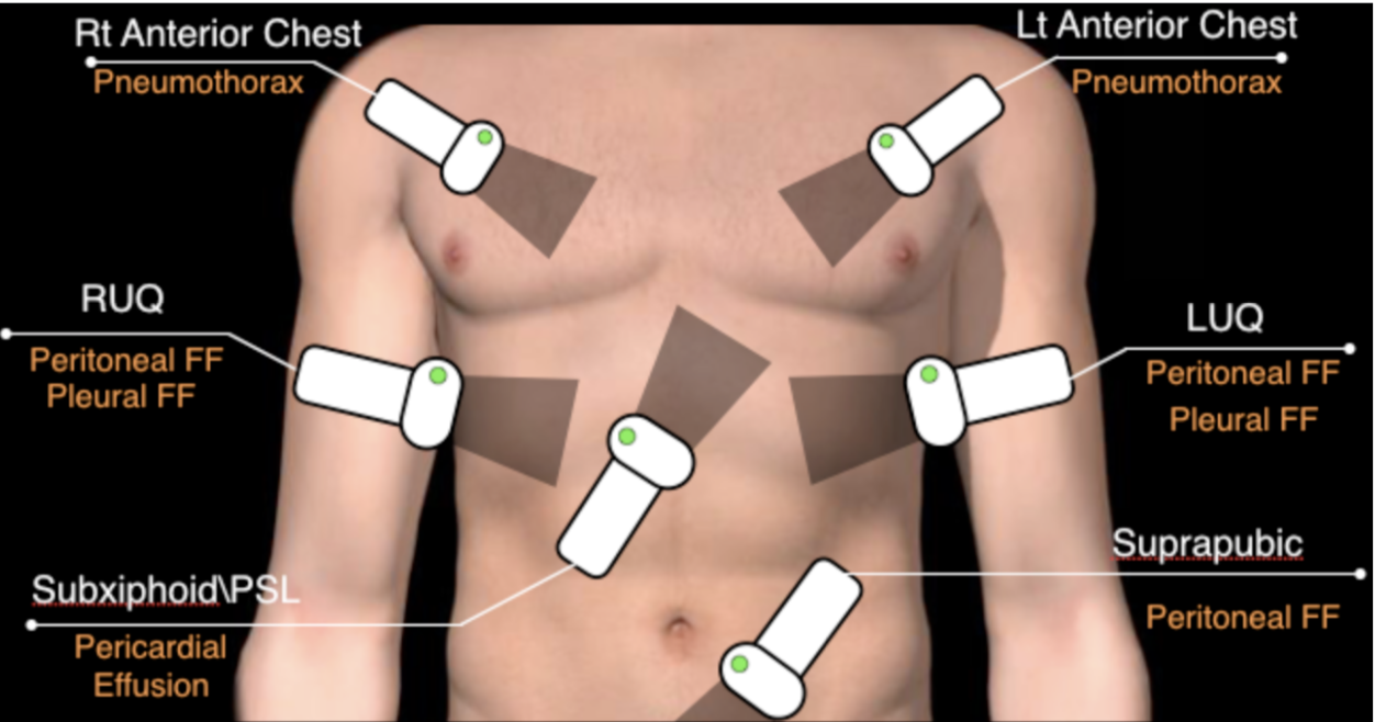
Probe position and normal images obtained during E-FAST examination.
Par un écrivain mystérieux
Last updated 07 juillet 2024

Download scientific diagram | Probe position and normal images obtained during E-FAST examination. (A) Right upper quadrant view demonstrating interface between liver and kidney (Morison's pouch). (B) Left upper quadrant view demonstrating spleen and kidney interface. (C) Left transverse view of the bladder. (D) Subcostal or subxiphoid view using the liver as a window to view the heart. (E) Anterior lung view. (F) Anterior lung view with US set to motion mode: this depicts a 1-dimensional view (marked on the top of the screen) as it changes over time (marked on the bottom of the screen); straight lines represent static soft tissue above the granular pattern representing the sliding of the pleura back and forth over time. E-FAST, Extended Focused Assessment with Sonography in Trauma examination. Used by permission from Introduction to Bedside Ultrasound, Vol 1, Dawson M, Mallin M, eds. Lexington, KY: Emergency Ultrasound Solutions; 2012: chap 1. from publication: Point-of-Care Ultrasound in Established Settings | The original and most widely accepted applications for point-of-care ultrasound (POCUS) are in the settings of trauma, shock, and bedside procedures. Trauma was the original setting for the introduction of POCUS and has been standardized under the four-plus view examination | Point-of-Care Systems, Ultrasound and Ultrasonography | ResearchGate, the professional network for scientists.

Focused Assessment with Sonography in Trauma (FAST) in 2017: What Radiologists Can Learn

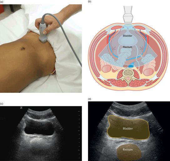
Focused Assessment with Sonography for Trauma (FAST) Exam: Image Acquisition

Focused Assessment with Sonography in Trauma (FAST) in 2017: What Radiologists Can Learn
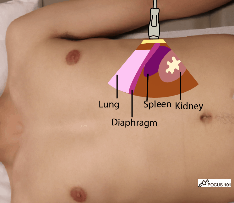
eFAST Ultrasound Exam Made Easy: Step-By-Step Guide - POCUS 101
FAST Sonoguide

Rectal examination - Wikipedia
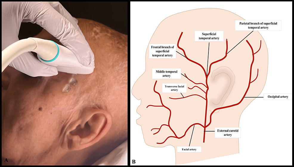
Cureus, Temporal Artery Ultrasound for the Diagnosis of Giant Cell Arteritis in the Emergency Department

The E-FAST Exam – Chapter 26 Media Library – WFUMB
The extended focused assessment with sonography for trauma (E-FAST)

FAST Sonoguide
EMERGENCY ULTRASOUND
Recommandé pour vous
 eFAST — MMHEME14 Jul 2023
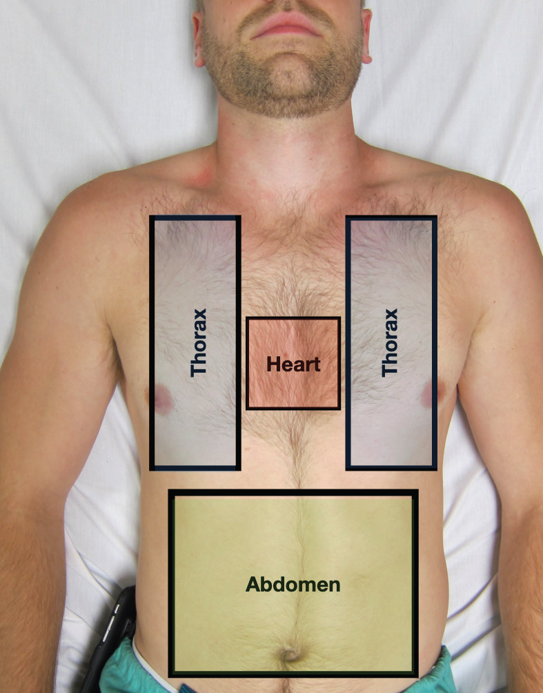 eFAST 2.0: Refining an Integral Trauma Exam - ACEP Now14 Jul 2023
 Focused assessment with sonography for trauma - Wikipedia14 Jul 2023
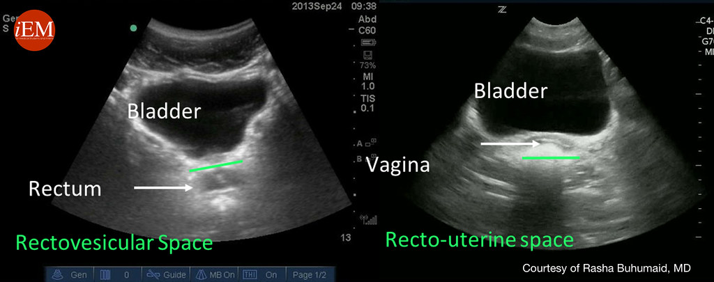 eFAST, Extended Focused Assessment using Sonography in Trauma14 Jul 2023
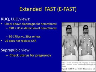 Fast Scan14 Jul 2023
 ▷ Utilidad del protocolo E-FAST en la Medicina de Urgencias y Emergencias - Ocronos - Editorial Científico-Técnica14 Jul 2023
- E-Fast14 Jul 2023
 Letter e fast speed logo Royalty Free Vector Image14 Jul 2023
 Ford Mustang Mach-E EV: A Road-Trip Fast-Charging Strategy Guide14 Jul 2023
 Protocolo EFAST en urgencias (Curso de ecografía en urgencias de la SEMFYC)14 Jul 2023

Tu pourrais aussi aimer
 PRYM VARIO Creative® Tool bei Schnuckidu14 Jul 2023
 Calculette image stock. Image du prévoir, numéroté, électronique14 Jul 2023
 Ramadan Mubarak Lune Led Cordon Lumières Ramadan Décorations pour Home Festival Guirlande Éclairage Eid Moubarak Décor Ramadan Kareem C14 Jul 2023
 LED Rope Lights Plug in Operated String Lights Hanging Fairy Lights with Remote for Camping Party Halloween Christmas Decoration, 30M-300LED14 Jul 2023
 Voitures Volkswagen Golf 7 d'occasion - Autohero14 Jul 2023
 iPhone 11 Pro Max Symmetry Series Clear Case14 Jul 2023
 Ventilateur 200mm Enthusiast Fan FS-200 - NZXT - Radiateurs14 Jul 2023
 8 300+ Panier à Linge Photos, taleaux et images libre de droits - iStock14 Jul 2023
 Clé valve universelle pour pneumatique et semi rigides toutes marques14 Jul 2023
 Jul - créateur de bd - Ma vie d'artiste14 Jul 2023